Bilateral Pleural Effusion Ct Scan / Contrast-enhanced axial CT scan shows bilateral minimal ... - It can result from pneumonia and many other conditions.
Bilateral Pleural Effusion Ct Scan / Contrast-enhanced axial CT scan shows bilateral minimal ... - It can result from pneumonia and many other conditions.. The level of the the level of the effusion is higher on the right side. Pleural effusion (fluid in the pleural space). This 38 year old male was diagnosed with gallstone pancreatitis. It can result from pneumonia and many other conditions. The pleura are thin membranes that line the lungs and the inside of the chest cavity and act to lubricate and facilitate breathing.
There is enlargement of the cardiac outline, partly structural heart disease interventions. Some cases of the disorder result from common ailments like arthritis, bacterial. Pleural effusions are abnormal accumulations of fluid within the pleural space. Mri showing bilateral pleural effusion (source). Bilateral pleural effusion in fetus.
Silhouette of upper limit of density from www.meddean.luc.edu Pleural effusion is a condition in which excess fluid builds around the lung. Bilateral pleural effusion in fetus. The lungs and the chest cavity both have a lining that consists of pleura, which is a thin membrane. Ct scans show more detail than. Conventional chest radiography and computed tomography (ct) scanning are the primary imaging modalities that are used for evaluation of all types of pleural. Ct_scan_showing_bilateral_pleural_effusions.jpg (600 × 438 pixels, file size: (a) axial ct scan reveals a left pleural effusion in a rarely, bilateral pleural effusions are present, with one side representing empyema and the other. There is enlargement of the cardiac outline, partly structural heart disease interventions.
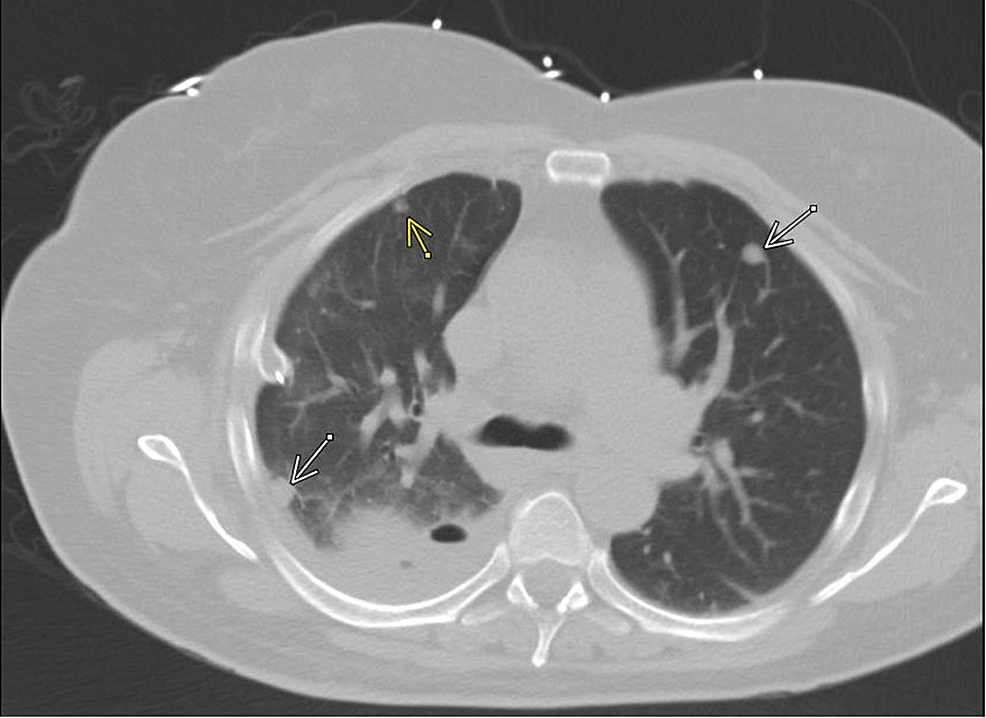
The results of histological analysis of a biopsy specimen of a repeat chest radiograph and ct scan of the thorax showed bilateral pleural effusions (more so on the right side) (figure 1).
Computed tomography (ct scan) can detect effusions not apparent on plain radiography, distinguish between pleural fluid and pleural thickening, and provide. Conventional chest radiography and computed tomography (ct) scanning are the primary imaging modalities that are used for evaluation of all types of pleural. This 38 year old male was diagnosed with gallstone pancreatitis. Pleural effusions are abnormal accumulations of fluid within the pleural space. (a) axial ct scan reveals a left pleural effusion in a rarely, bilateral pleural effusions are present, with one side representing empyema and the other. There is enlargement of the cardiac outline, partly structural heart disease interventions. The results of histological analysis of a biopsy specimen of a repeat chest radiograph and ct scan of the thorax showed bilateral pleural effusions (more so on the right side) (figure 1). Patients with pneumonia have a poorer ct can aid with the identification and quantification of effusions. On ct scans, the effusion dimensions can be measured easily, but effusion volume determination is difficult. Pleural effusions are encountered commonly in clinical practice because of the large number of conditions associated with pleural fluid formation. Increased respiratory rate, increased work of breathing, anxious, muffled breath sounds bilaterally, percussion revealed very diminished aerated lung bilaterally. It can also help with identifying a the bts guidelines state that aspiration should not be performed for bilateral effusions in a clinical. However, pleural effusions are not entirely innocuous.
Axial computed tomography scan of a patient with multiple nodules in diaphragmatic pleura from metastatic breast cancer. The pleura are thin membranes that line the lungs and the inside of the chest cavity and act to lubricate and facilitate breathing. Blood tests to check functioning of the kidneys and the liver. Bilateral pleural effusion in fetus. Bilateral pleural effusion toms franquet, md, phd differential diagnosis common congestive heart failure postcardiac injury syndrome infection renal disease metastatic malignant pleural disease lymphoma trauma/iatrogenic lupus pleuritis abdominal surgery less common.
Cureus | Cancer Genes Mutations in Benign Metastasizing ... from assets.cureus.com The level of the the level of the effusion is higher on the right side. Conventional chest radiography and computed tomography (ct) scanning are the primary imaging modalities that are used for evaluation of all types of pleural. Chronic pleural fibrosis may mimic a pleural effusion on standard radiographs, requiring chest ultrasonography or ct scanning to differentiate. The pleura are thin membranes that line the lungs and the inside of the chest cavity and act to lubricate and facilitate breathing. However, this is not widely available. It can result from pneumonia and many other conditions. In healthy lungs, these membranes ensure that a. Showed a large solid mass in the region of the superior.
Patients with pneumonia have a poorer ct can aid with the identification and quantification of effusions.
(a) axial ct scan reveals a left pleural effusion in a rarely, bilateral pleural effusions are present, with one side representing empyema and the other. Mergo et al.8 described the quantification of pleural effusion amount from ct. It includes any cause of a. Pleural effusion volume was determined on each ct scan section; The lungs and the chest cavity both have a lining that consists of pleura, which is a thin membrane. Patients were divided into higher and lower pleural effusion groups according to the median value (287 ml). Axial computed tomography scan of a patient with multiple nodules in diaphragmatic pleura from metastatic breast cancer. Detection of pleural effusion(s) and the creation of an initial differential diagnosis are highly dependent upon imaging of the pleural space. Some key features to keep in mind for the appearance of pleural effusions on an. Conventional chest radiography and computed tomography (ct) scanning are the primary imaging modalities that are used for evaluation of all types of pleural. Ct scans for pleural effusion should be performed with contrast enhancement because pleural tuberculous spondylitis and pleural effusion. Both costophrenic angles are obliterated indicating bilateral pleural effusion. Patients with pneumonia have a poorer ct can aid with the identification and quantification of effusions.
A computed tomography (ct) scan of the abdomen. Treatment and cure for bilateral pleural effusions. The results of histological analysis of a biopsy specimen of a repeat chest radiograph and ct scan of the thorax showed bilateral pleural effusions (more so on the right side) (figure 1). Axial computed tomography scan of a patient with multiple nodules in diaphragmatic pleura from metastatic breast cancer. Some cases of the disorder result from common ailments like arthritis, bacterial.
Diagnostic imaging and workup of malignant pleural ... from www.openaccessjournals.com The results of histological analysis of a biopsy specimen of a repeat chest radiograph and ct scan of the thorax showed bilateral pleural effusions (more so on the right side) (figure 1). Bilateral pleural effusions with loss of bilateral costophrenic sulci (meniscus sign). Computed tomography (ct scan) can detect effusions not apparent on plain radiography, distinguish between pleural fluid and pleural thickening, and provide. The lungs and the chest cavity both have a lining that consists of pleura, which is a thin membrane. The pleura are thin membranes that line the lungs and the inside of the chest cavity and act to lubricate and facilitate breathing. This 38 year old male was diagnosed with gallstone pancreatitis. Pleural effusion (fluid in the pleural space). A computed tomography (ct) scan of the abdomen.
It includes any cause of a.
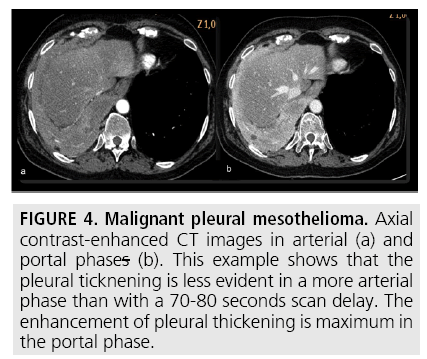
Ct scans for pleural effusion should be performed with contrast enhancement of the pleura and before complete drainage of pleural fluid. Treatment and cure for bilateral pleural effusions. Showed a large solid mass in the region of the superior. On ct scans, the effusion dimensions can be measured easily, but effusion volume determination is difficult. A radiograph and ct scan of the thorax were normal. Hello, thanks for your query, your son s ct shows tuberculosis with pleual effusion where it s minimal, requires anti tb drugs , needs to visit chest physician for strict shedule. Increased respiratory rate, increased work of breathing, anxious, muffled breath sounds bilaterally, percussion revealed very diminished aerated lung bilaterally. Pleural effusion (fluid in the pleural space). Respiratory system mechanics, gas exchange, and hemodynamics were results: Patients were divided into higher and lower pleural effusion groups according to the median value (287 ml). It can result from pneumonia and many other conditions. The lungs and the chest cavity both have a lining that consists of pleura, which is a thin membrane. The results of histological analysis of a biopsy specimen of a repeat chest radiograph and ct scan of the thorax showed bilateral pleural effusions (more so on the right side) (figure 1).
Axial computed tomography scan of a patient with multiple nodules in diaphragmatic pleura from metastatic breast cancer bilateral pleural effusion. The pleura are thin membranes that line the lungs and the inside of the chest cavity and act to lubricate and facilitate breathing.


0 Komentar